![]() Figure 2 of
Lin, Mol Vis 2007;
13:1822-1827.
Figure 2 of
Lin, Mol Vis 2007;
13:1822-1827.
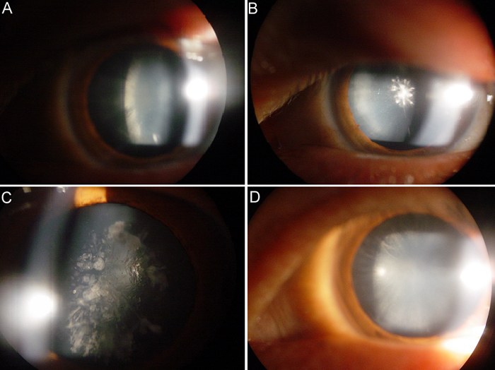
Figure 2. Binocular slit lamp photographs of the affected individuals
A: The right eye of individual IV:9 shows fine punctate opacities in the posterior cortex. B: The left eye of individual IV:9 shows the anterior polar cataract with punctate opacities in the anterior cortex. C: The right eye of individual IV:7 shows fine punctate opacities in the cortex and core of the lens. D: The left eye of individual IV:7 shows a mass of irregular opacification clustering in the anterior cortex.