![]() Figure 3 of
Redmond, Mol Vis 2007;
13:1813-1821.
Figure 3 of
Redmond, Mol Vis 2007;
13:1813-1821.
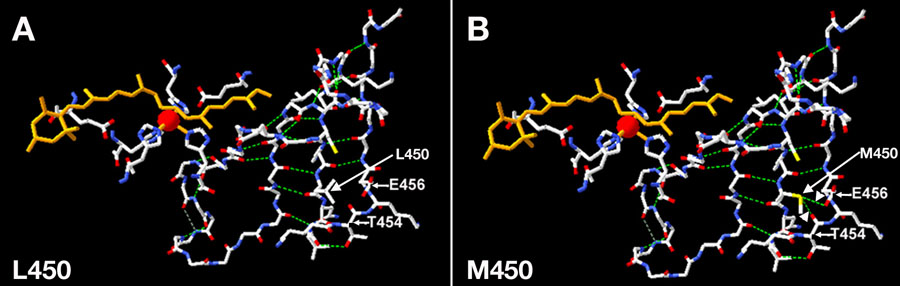
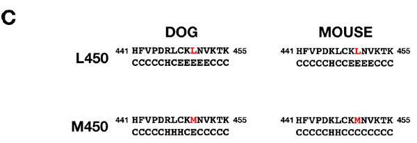
Figure 3. Effect of changing leucine at aa450 to methionine on predicted tertiary structure of RPE65, modeled on ACO, and on secondary structure prediction of RPE65
A and B: Mouse RPE65 residues are plotted onto the ACO backbone [26] using DeepView. A: L450 (wildtype) showing lack of interaction of L450 with other residues; B: M450 showing interaction of M450 with T454 and E456. Arrowheads indicate extra side-chain H-bonding due to M450. C: Effect of M450 mutation on predicted secondary structure of RPE65. M450 abolishes predicted β-strand number 26 in both dog and mouse RPE65. E indicates predicted extended β-strand, H indicates α-helix, and C indicates other structure.