![]() Figure 1 of
Redmond, Mol Vis 2007;
13:1813-1821.
Figure 1 of
Redmond, Mol Vis 2007;
13:1813-1821.
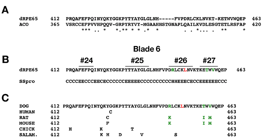
Figure 1. Sequence analysis of Blade 6 of RPE65
A: Alignment of the aa412-aa463 region of RPE65 with Blade 6 region of ACO (aa365-aa420); Asterisk indicates identity and dot indicates similarity. B: SSpro secondary structure prediction of the aa412-aa463 region of RPE65; numbers above sequence indicate corresponding β-strand number from apocarotenoid oxygenase predicted structure; E indicates predicted extended β-strand, H indicates α-helix, and C indicates other structure. C: Alignment of the aa412-aa463 region of dog, human, rat, mouse, chicken, and salamander RPE65 showing position of residue 450 (red L), and of variant residues of rodent RPE65 in β-strand number 26 and number 27.