![]() Figure 2 of
Mott, Mol Vis 2007;
13:1802-1812.
Figure 2 of
Mott, Mol Vis 2007;
13:1802-1812.
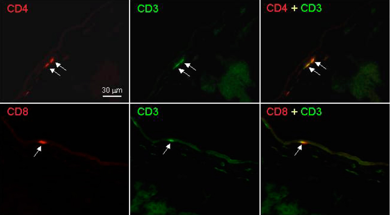
Figure 2. Detection of CD4/CD3-postitive and CD8/CD3-positive T cells in immunostained sections of the cornea of naive mice
Representative corneal sections from naive BALB/c mice stained with anti-CD4 and anti-CD3 or anti-CD8 and anti-CD3 mAbs as described in Methods are shown. Panels: Sections of cornea from naive BALB/c mice were stained with anti-CD4 (CD4), anti-CD3 (CD3), or merged (CD4+CD3). Arrows show CD4+, CD3+, and CD4/CD3 double-positive T cells in the stromal cell layer of the cornea. Sections of the cornea from naive BALB/c mice were stained with anti-CD8 (CD8), anti-CD3 (CD3), or merged (CD8/CD3). The arrows show CD8+, CD3+, and CD8/CD3 double-positive T cells in the stromal cell layer of the cornea.