![]() Figure 1 of
Mott, Mol Vis 2007;
13:1802-1812.
Figure 1 of
Mott, Mol Vis 2007;
13:1802-1812.
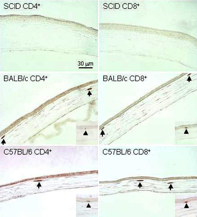
Figure 1. Detection of CD4+ and CD8+ T cells in immuostained sections of the cornea of naive mice by light microscopy
Representative corneal sections from female, eight-week-old naive BALB/c, C57BL/6, and SCID mice stained with anti-CD4 and anti-CD8 antibodies as described in Methods are shown. Panels: Sections of cornea from naive SCID mice were stained with anti-CD4 (SCID CD4+) or anti-CD8 antibody (SCID CD8+). No CD4+ or CD8+ T cells were observed in any of the examined sections. Sections of cornea from naive BALB/c mice were stained with anti-CD4 (BALB/c CD4+) or anti-CD8 antibody (BALB/c CD4+). The arrows show CD4+ and CD8+ T cells in the stroma and epithelial cell layers (insets) of the cornea. Sections of cornea from naive C57BL/6 mice were stained with anti-CD4+ (C57BL/6 CD4+) or anti-CD8+ antibody (C57BL/6 CD8+). The arrows show CD4+ and CD8+ T cells in the stroma and epithelial cell layers (insets) of the cornea.