![]() Figure 2 of
Dekomien, Mol Vis 2007;
13:174-180.
Figure 2 of
Dekomien, Mol Vis 2007;
13:174-180.
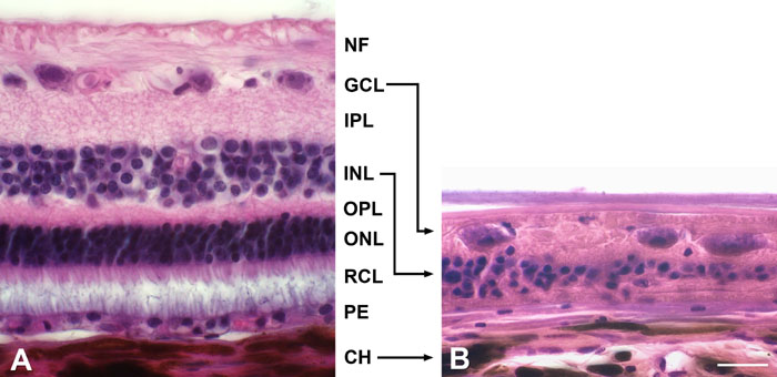
Figure 2. Hematoxylin-, eosin-stained paraffin sections illustrating normal canine retina and gPRA-affected retina of a Schapendoes dog
A: The retina of a Saarloos/Wolfshound exhibited regular nuclear layers with the nerve fiber layer (NF), ganglion cell layer (GCL), inner plexiform layer (IPL), inner nuclear layer (INL), outer plexiform layer (OPL), outer nuclear layer (ONL), photoreceptor layer (RCL), and the pigment epithelium (PE). B: In the degenerated retina of a Schapendoes dog the outer retina with the RCL and ONL was missing, the INL and the IPL reduced, the GCL was comparatively preserved. CH represents choroid. The scale bar in B represents 20 μm; same magnification for A and B.