![]() Figure 5 of
Jiang, Mol Vis 2007;
13:1783-1792.
Figure 5 of
Jiang, Mol Vis 2007;
13:1783-1792.
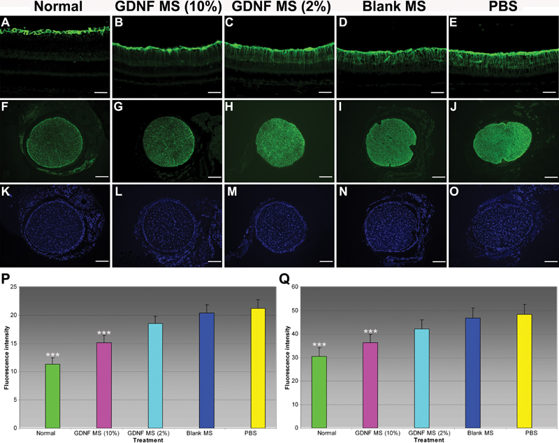
Figure 5. GDNF microspheres decreased GFAP expression of retina and optic nerve
GFAP expression was mainly localized to the inner limiting membrane in normal retina (A). Chronic IOP elevation resulted in increased GFAP expression (B-E). Scale bars represent 100 μm. Chronic IOP elevation increased the GFAP expression in an ON cross section (G-J) compared with that of normal ON without IOP elevation (F). Ten percent of GDNF microsphere (MS) treatment (G) decreased the GFAP expression, and 2% GDNF MS treatment (H) moderately decreased the GFAP expression compared with blank MS (I) and PBS treatment (J). K-O illustrate the corresponding optic nerve section stained with DAPI. Scale bars represent 200 μm. Quantitative analysis of GDNF MS treatment on the GFAP expression of retina (P) and ON (Q). Chronic IOP elevation resulted in significantly increased GFAP expression in the retina (p<0.001) and optic nerve (p<0.001) compared with that of normal tension eyes. Ten percent of GDNF microspheres significantly decreased the IOP-induced GFAP overexpression in both the inner retina (p<0.001) and ON (p<0.001) while 2% GDNF microspheres were more difficult to distinguish from treatment with blank microspheres and PBS alone (p>0.05). Three asterisks indicate p<0.001. In the figure, MS represents microsphere.