![]() Figure 1 of
Yellore, Mol Vis 2007;
13:1777-1782.
Figure 1 of
Yellore, Mol Vis 2007;
13:1777-1782.
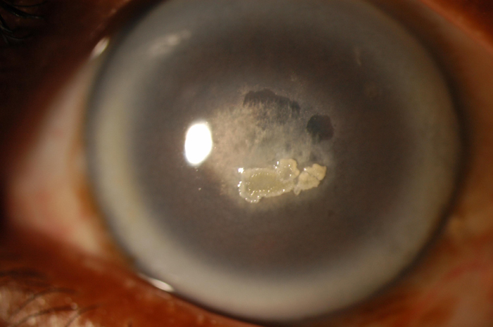
Figure 1. Schnyder crystalline corneal dystrophy
Slit lamp photomicrograph of the proband from a previously unreported family with SCCD demonstrates dense arcus lipoides and central corneal opacification secondary to superficial and subepithelial crystalline deposits. Photograph courtesy of Dr. Sadeer Hannush.