![]() Figure 2 of
Li, Mol Vis 2007;
13:1758-1768.
Figure 2 of
Li, Mol Vis 2007;
13:1758-1768.
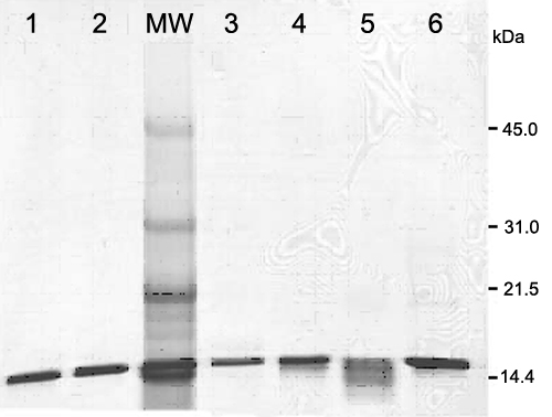
Figure 2. SDS-PAGE analysis of D51 αA-crystallin and mutants
Samples were isolated and purified as described in Methods. Coomassie blue staining revealed the positions of molecular weight standards (MW), D51-GGG (lane 1), D51-GPG (lane 2), D51-TPT (lane 3), D51-IGV (lane 4), D51-ITV (lane 5), and D51-αA-crystallin (lane 6) after purification.