![]() Figure 3 of
Valamanesh, Mol Vis 2007;
13:1746-1757.
Figure 3 of
Valamanesh, Mol Vis 2007;
13:1746-1757.
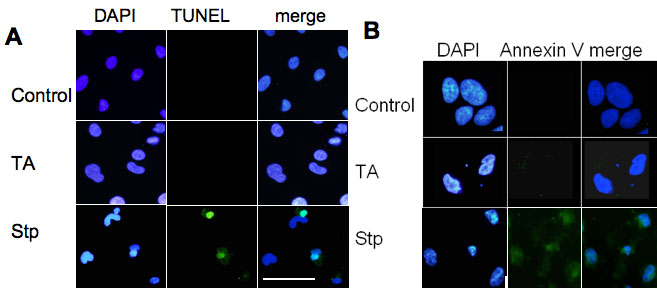
Figure 3. Absence of apoptosis markers in ARPE-19 cells treated with triamcinolone acetonide
Subconfluent ARPE 19 cells were incubated for 72 h in the presence triamcinolone (TA). Cells were then stained with TUNEL assay (A) or with annexin V (B). ARPE cells induced to die by treatment with 1 μM staurosporin (stp) for 24 h were used as positive controls (stp). Scale bar represents 50 μm.