![]() Figure 1 of
Valamanesh, Mol Vis 2007;
13:1746-1757.
Figure 1 of
Valamanesh, Mol Vis 2007;
13:1746-1757.
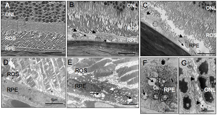
Figure 1. Structural alterations of the rat retina 8 days after intravitreous injection of Kenacort
A: Retina from a PBS-treated rat (control). B, C: Retina from Kenacort®-injected eyes. Thin black arrows show vacuoles and enlarged retinal pigment epithelium (RPE) cells. Thick arrows indicate increased RPE microvilli length. Scale bar represents 10 μm. Lower panels are ultrathin sections. D: Retina from a PBS-treated rat (control). E, F, G: Retina from Kenacort®-injected eyes. E: RPE cells show cytosolic vacuoles and dilated mitochondria (black arrows), as well as undigested debris (white arrow). A higher magnification of degraded mitochondria is shown on F (arrows). G: Vacuoles (arrows) are also observed in retina glial Müller cells prolongations. Note preservation of photoreceptor nuclei. ROS indicates rod outer segments, ONL indicates outer nuclear layer.