![]() Figure 2 of
Yamanaka, Mol Vis 2007;
13:1730-1739.
Figure 2 of
Yamanaka, Mol Vis 2007;
13:1730-1739.
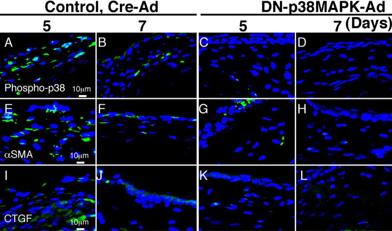
Figure 2. Expression of phospho-p38, αSMA and CTGF in mouse mechanical injured conjunctiva treated with either control Cre-Ad or DN-p38MAPK-Ad
The epithelium and mesenchymal cells in the injured conjunctiva are labeled with anti-phospho-p38MAPK antibody markedly on day 5 and faintly on day 7 (A,B), whereas those in the DN-p38MAPK-Ad group specimens are not, or just faintly, stained (C,D). Immunohistochemical analysis of alpha SMA shows that many fibroblasts in the subconjunctiva are labeled with anti-αSMA antibody in the control Cre-Ad specimens, but a few fibroblasts in DN-p38MAPK-Ad specimens were labeled on day 5 (E,G). On day 7, a few cells were still positive in the DN-p38MAPK-Ad group, but not in the control (F,H). The expression pattern of CTGF is similar to that of αSMA (I-L). The scale bar is equal to 10 μm.