![]() Figure 1 of
Yamanaka, Mol Vis 2007;
13:1730-1739.
Figure 1 of
Yamanaka, Mol Vis 2007;
13:1730-1739.
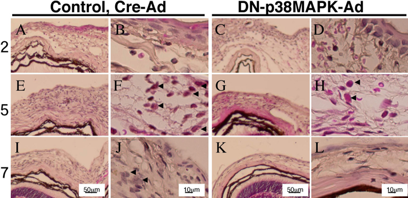
Figure 1. Histology of mouse mechanical injured conjunctiva treated with either control Cre- or DN-p38MAPK-Ad
HE staining shows that DN-p38MAPK introduction also apparently suppresses the conjunctival edema and inflammatory cell infiltration as compared to the Cre-Ad group (A-L). HE staining also shows that epithelial closure of the mechanical incision in the mouse conjunctiva seemed to be facilitated with topical application of adenoviral vector of DN- p38MAPK at day 5 (E-H). On day 7 in both the control and treatment groups, the conjunctival epithelium is resurfaced (I-L). Arrow head; Polymorphonuclear leukocytes were seen in subconjunctival tissue at day 2 -5, but there seemed no difference of the distribution of this cell type. The scale bar is equal to 50 μm in A, C, E, G, I, and K, and equal to 10 μm in B, D, F, H, J, and L.