![]() Figure 2 of
Baharvand, Mol Vis 2007;
13:1711-1721.
Figure 2 of
Baharvand, Mol Vis 2007;
13:1711-1721.
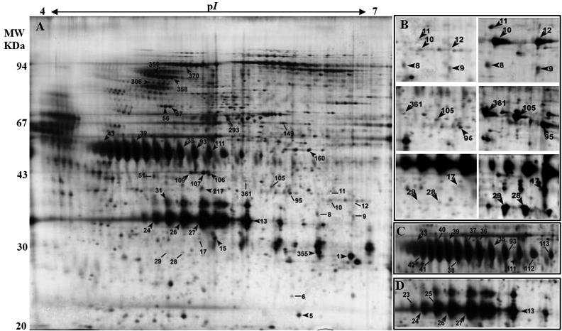
Figure 2. 2-DE gel analysis of epithelium-denuded amniotic membrane proteins
2-DE gel analysis of proteins extracted from epithelium-denuded amniotic membrane. The first dimension was performed using 120 μg total soluble proteins on linear gradient IPG strips with a pH of 4-7. In the second dimension, 12% SDS-PAGE gels were used, and proteins were visualized using silver nitrate (A). The arrows and lines show identified proteins with similar (arrows) and different (lines) expression patterns across six individuals. Examples of changes in protein abundance in different individuals (B) and different forms of lumican (C) and osteoglycin (D) on 2-DE gels have been presented.