![]() Figure 1 of
Baharvand, Mol Vis 2007;
13:1711-1721.
Figure 1 of
Baharvand, Mol Vis 2007;
13:1711-1721.
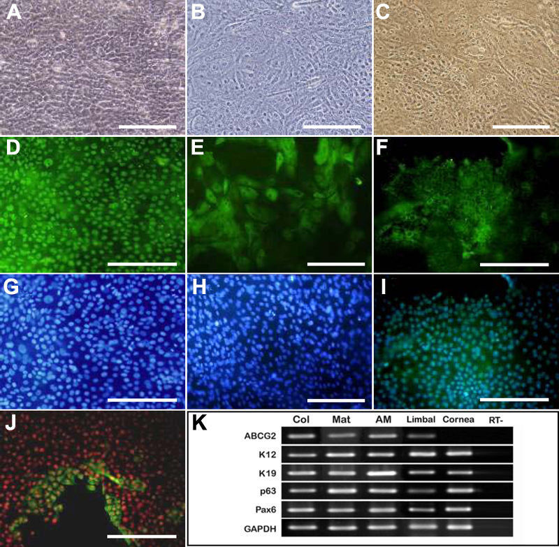
Figure 1. Human limbal epithelium from limbal explants expanded on different extracellular matrices at day 14
Phase contrast microscopy of the cells on AM (A), matrigel (B), and collagen (C) are shown. Representative immunofluorescent staining of the cells on AM with corresponding nucleus counterstaining by Hoechst 33342 (blue) and propidium iodide (red). (D) Staining of p63 in nucleus, (E) staining of K19 in cytoplasm, (F) staining of Cx43 in the cell membrane, (G) staining of K3 in cytoplasm. Scale bar: 200 μm. (H) mRNA expression of proposed markers in normal limbus and cornea and cultured cells on different ECMs.