![]() Figure 2 of
Roman, Mol Vis 2007;
13:1701-1710.
Figure 2 of
Roman, Mol Vis 2007;
13:1701-1710.
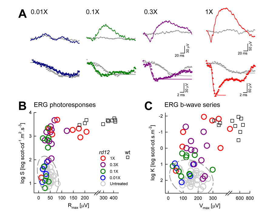
Figure 2. Electroretinography parameters of rd12 mice to different doses of subretinal AAV2-hRPE65
A: ERGs evoked by 0.1 log scot-cd.s.m-2 flashes (upper row) and by 3.6 log scot-cd.m.s-2 flashes (lower row) in treated (colors) and untreated (gray) eyes of one rd12 mouse from each dose group. As vector dose increases, responses become asymmetric with treated retinas showing increasing amplitude of b-waves and faster photoresponses. Photoreceptor activation models (smooth lines) fit to the photoresponses are shown. All traces start at stimulus onset. B: Photoresponse parameters in rd12 eyes treated with a range of vector doses. As dosage increases above 0.01X, parameter pairs drift outside of the 99% confidence region (dashed ellipse) defined by the untreated eyes of rd12 animals and start approaching wt levels. C: Luminance response parameters in treated rd12 eyes similarly show a dose-related progression from the region corresponding to untreated eyes to the region corresponding to wt eyes.