![]() Figure 1 of
Roman, Mol Vis 2007;
13:1701-1710.
Figure 1 of
Roman, Mol Vis 2007;
13:1701-1710.
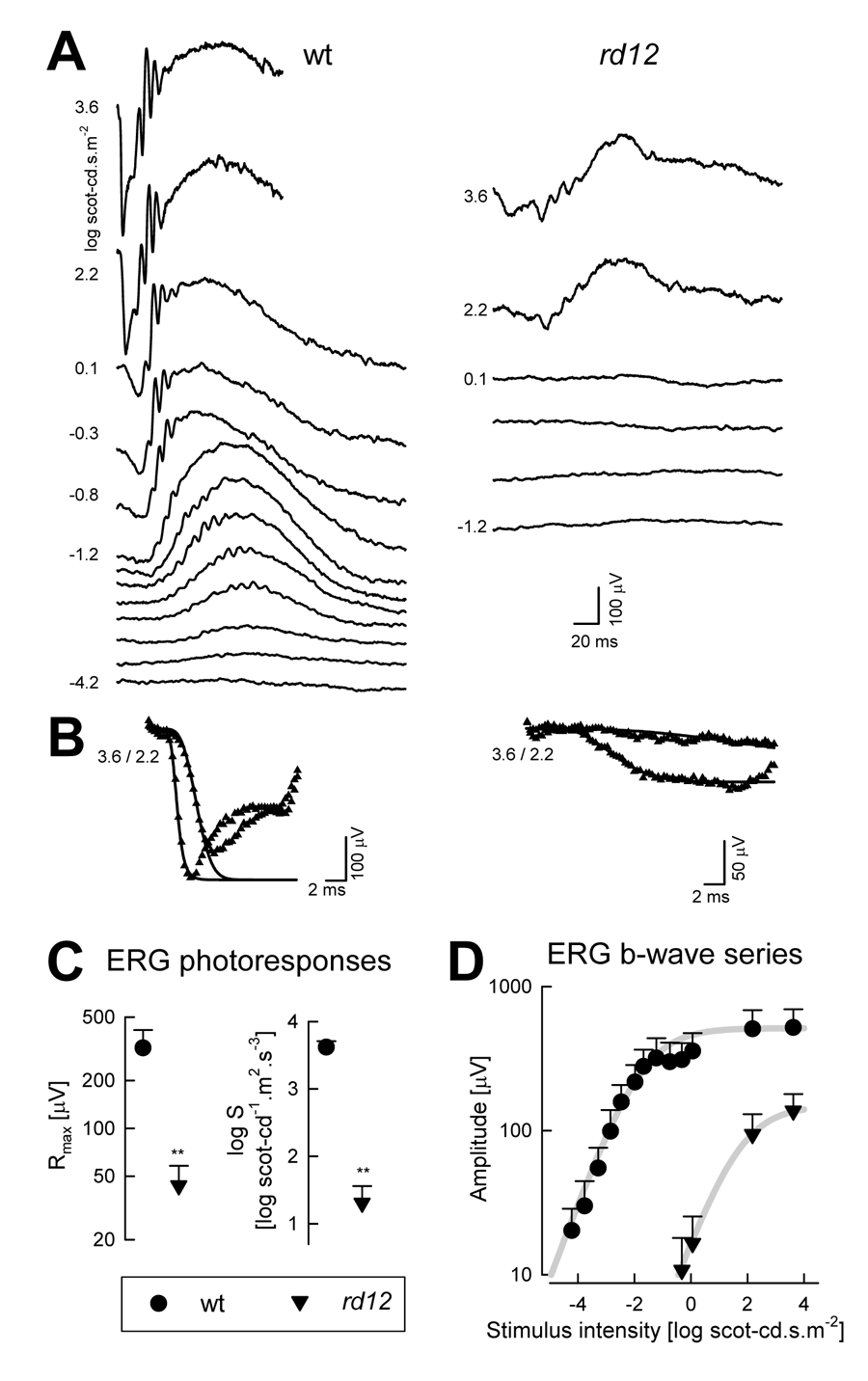
Figure 1. Definition of electroretinography abnormalities in the rd12 mouse model of RPE65-LCA
A: Dark adapted ERGs to increasing stimulus intensities (shown to the left of key traces) for representative 2-month-old wt and rd12 mice. Blue flashes were used for all intensities except the highest, which were evoked by white flashes. Traces start at stimulus onset. B: ERG photoresponses (symbols) evoked by 3.6 and 2.2 log scot-cd.s.m-2 flashes are fit as an ensemble with a model of phototransduction (smooth lines). The response from the mutant shows reduced amplitude and sensitivity. C: Summary statistics of maximum amplitude (Rmax) and sensitivity (log S) parameters obtained from photoresponse modeling in rd12 mice are significantly (*) different than wt. D: Luminance-response functions derived from ERG b-wave series show diminished light sensitivity in mutant animals indicated by a shift to the right of the curves. Mutant animals also show a reduction in maximum amplitude. Error bars equals to 1SD.