![]() Figure 4 of
Correa-Gomez, Mol Vis 2007;
13:1695-1700.
Figure 4 of
Correa-Gomez, Mol Vis 2007;
13:1695-1700.
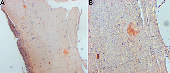
Figure 4. Histopathological features of an affected cornea carrying the A546D TGFBI mutation
Light microscopy of excised corneal tissue stained with congo red revealed variably-sized stromal deposits of amyloid material, predominantly in the mid and posterior stroma. A: 10X; B: 40X