![]() Figure 2 of
Correa-Gomez, Mol Vis 2007;
13:1695-1700.
Figure 2 of
Correa-Gomez, Mol Vis 2007;
13:1695-1700.
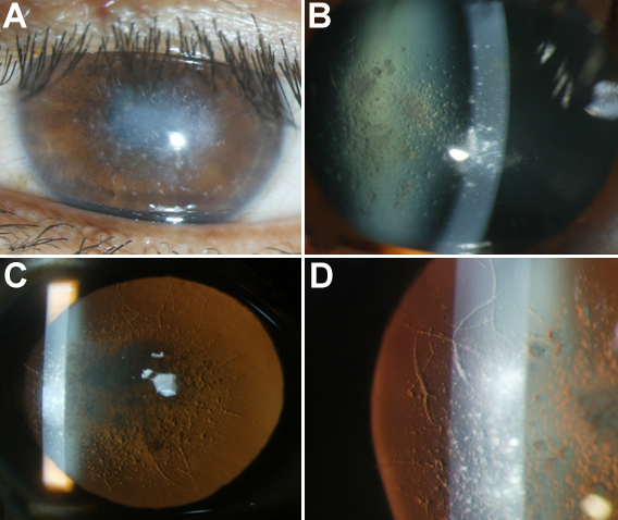
Figure 2. Corneal phenotype associated with the A546D TGFBI mutation
Slit lamp photographs of the proband show significant stromal opacification in the central part of the cornea (A and B). Corneal retroillumination demonstrated numerous central lesions combined with multiple linear opacities in central and peripheral cornea producing a typical lattice pattern (C). Small granular opacities are also evident in the periphery (D).