![]() Figure 2 of
Nezzar, Mol Vis 2007;
13:1641-1650.
Figure 2 of
Nezzar, Mol Vis 2007;
13:1641-1650.
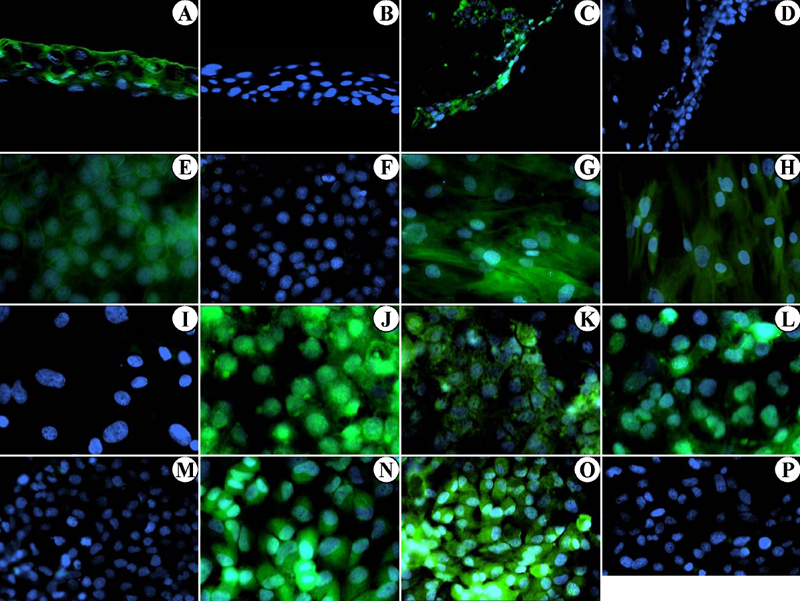
Figure 2. Immunolabeling of nuclear receptor, retinoin acid receptor, and retinoid X receptor proteins in human corneal and conjunctival cells and tissues
RAR α (A, J, N), RAR β (E, L), RAR γ (G, K, O), and RXR α (C, H) immunolocalizations (green fluorescence staining) were performed in total cornea (A, B), total conjunctiva (C, D), corneal epithelial cells (E, F), corneal keratinocytes (G, H, I), conjunctival corneal cells (J, K, L, M), and conjunctival cells (N, O, P). Cell nuclei were visualized with DAPI staining (blue fluorescence; A-P). Negative controls for the 4 antibodies used are presented in B, D, F, I, M, and P. Acquisitions were made under a standard Axiophot fluorescence microscope (Zeiss). Magnifications for A to D were x200, and for E to P were x400.