![]() Figure 1 of
Soest, Mol Vis 2007;
13:1608-1617.
Figure 1 of
Soest, Mol Vis 2007;
13:1608-1617.
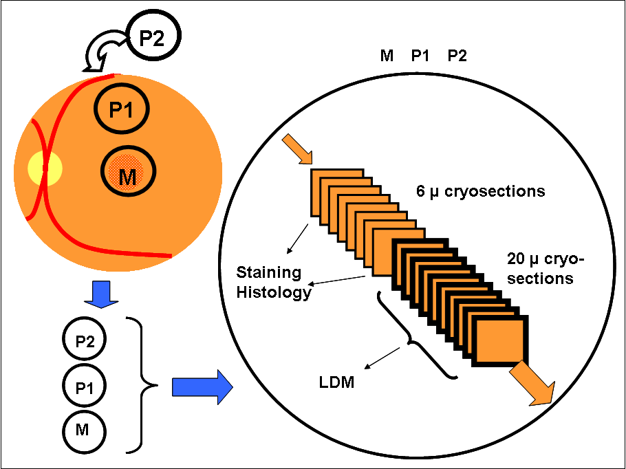
Figure 1. Strategy used for isolation of retinal pigment epithelium cells from cryo-sections from defined retinal locations
Top left: Shown are the topographical location of regions used in this study, where M represents macula, P1 represents paramacular area, P2 represents mid-postequatorial periphery. Bottom left: From freshly frozen and selected donor eyes, tissue fragments of 16 mm2 from each region (M, P1, P2) were cut out. Right: Serial cryo-sections were cut throughout the entire tissue fragment. Alternating series of eight 6 μm sections, for direct histological and (future) immunochemical stainings, and eight 20 μm sections for laser dissection microscopy (LDM) and RNA isolation, were cut.