![]() Figure 9 of
Bai, Mol Vis 2007;
13:1589-1600.
Figure 9 of
Bai, Mol Vis 2007;
13:1589-1600.
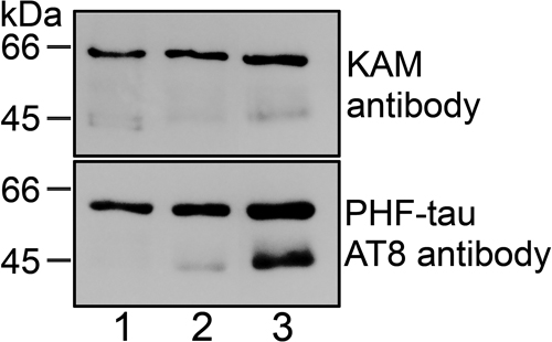
Figure 9. Immunoblot analysis of tau in mouse brain
Brain MAPs from 8-month-old wild type, αA-/--, and αB-/-- mice were prepared and the proteins were separated by SDS-PAGE. Samples were heated to 85 °C in SDS-PAGE sample buffer. Ten μg proteins were applied to each lane. Immunoblot analysis was performed with the monoclonal antibody KAM-MA305 to full length brain tau protein and the PHF-tau antibody AT-8. A: Wild type (Lane 1), αA-/-- (Lane 2), and αB-/-- (Lane 3) brain MAPs. Note that three tau-immunoreactive bands were readily detected in the wild type brains with the KAM-MA305 antibody, but the smallest peptide was undetectable in the αA-/-- and αB-/-- brains. B: Wild type (Lane 1), αA-/-- (Lane 2), and αB-/-- (Lane 3) brain MAPs. Tau-immunoreactive bands were detected in the wild type brains with the PHF-tau AT-8 antibody. Note that the only the largest immunoreactive peptide was strongly stained in the wild type brain, but the immunoreactivity of the PHF-tau AT-8 antibody to the smaller tau peptide increased significantly in the αA-/-- and αB-/-- over wild type samples.