![]() Figure 5 of
Bai, Mol Vis 2007;
13:1589-1600.
Figure 5 of
Bai, Mol Vis 2007;
13:1589-1600.
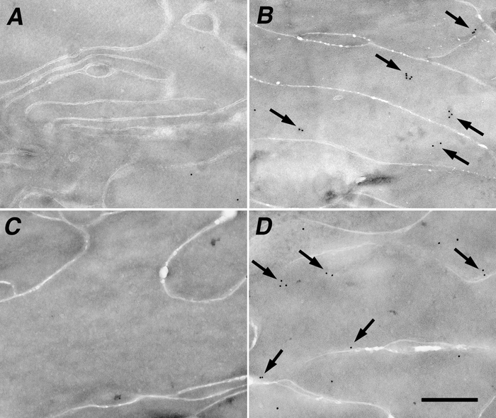
Figure 5. Cryo-immuno electron microscopy of tau in the lens
Anti-tau cryo-immuno electron microscopy in lens sections from 8-month-old mice with the antibody KAM-MA305 (A and B) or Tau-5 (C and D). A: Absence of cryo-immuno labeling in the lens cortical fiber cells of an 8-month-old wild type mouse. B: αB-/-- lens shows ample immunostaining with the tau antibody KAM-MA305, with clusters of immunogold particles in electron dense areas of the cells (arrows). C: Absence of immunostaining in 7-month-old wild type lens. D: αB-/-- lens shows ample immunostaining with the tau-5 antibody, with numerous immunogold particles in lens fiber cells (arrows). All images are shown at the same magnification. The scale bar is equal to 0.5 μm.