![]() Figure 4 of
Bai, Mol Vis 2007;
13:1589-1600.
Figure 4 of
Bai, Mol Vis 2007;
13:1589-1600.
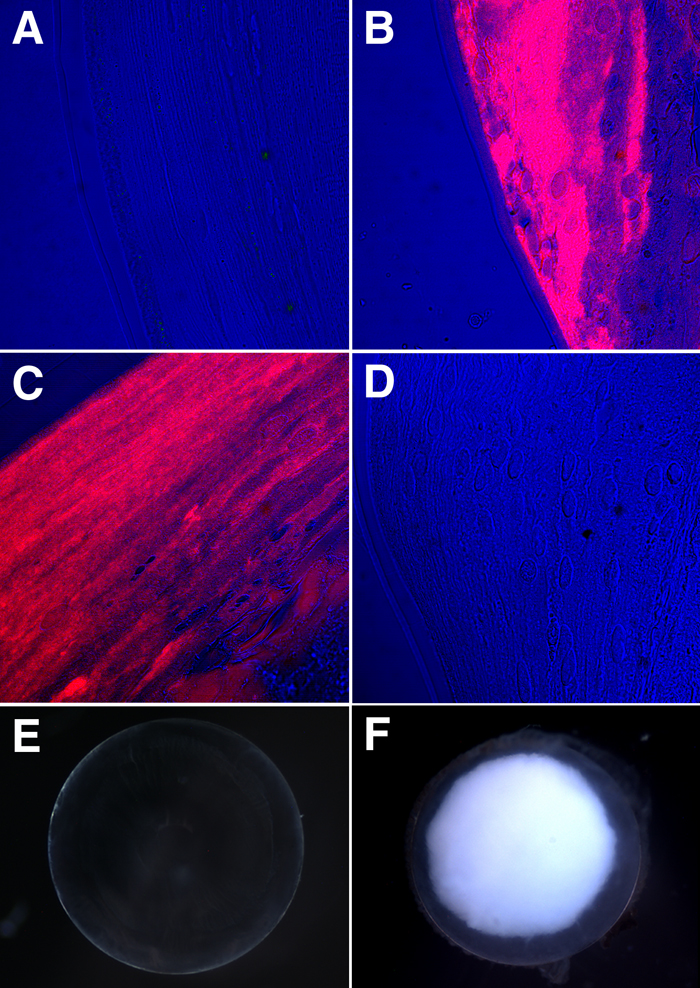
Figure 4. Immunofluorescence analysis of tau in the lens
Anti-tau immunostaining in lens sections from 6-month-old-mice. Red=tau; blue=DIC. A: Wild type lens. B: αA/B-/-- lens. C: αB-/-- lens. D: Absence of immunofluorescence in the lens specimen of a 6-month-old αA/B-/-- lens probed with non-immune mouse IgG. The scale bar is equal to 25 μm. E: Dark field image of a 6-month-old wild type mouse lens. F: Dark field image of a 6-month-old αA/B-/-- lens. Note that the opacity occupies three quarters of the lens. Note also that the outer cortex of the αA/B-/-- lens remains relatively clear.