![]() Figure 10 of
Bai, Mol Vis 2007;
13:1589-1600.
Figure 10 of
Bai, Mol Vis 2007;
13:1589-1600.
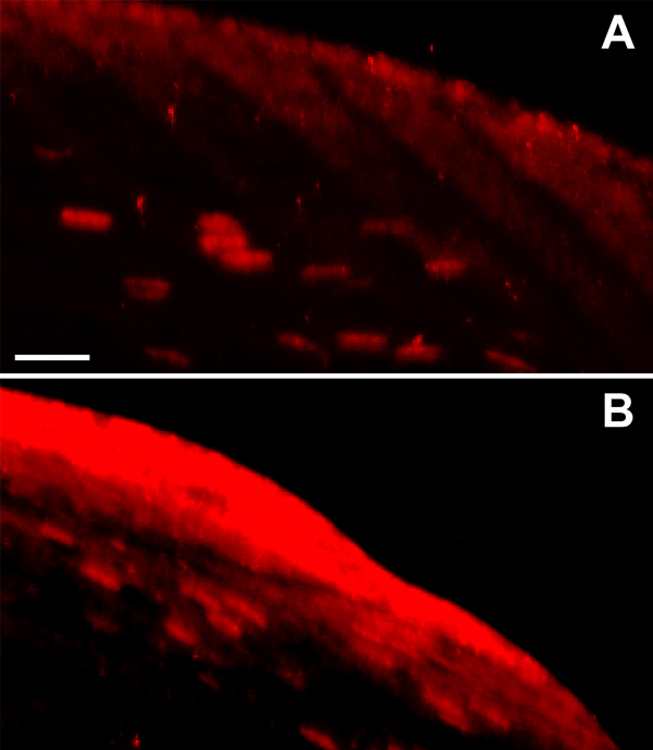
Figure 10. Expression of tau immunoreactivity in UVB mouse cataract model
Shown are the untreated lens (A) and the UVB-treated lens (B). Note the increase in lens cortical fiber cell immunostaining of tau in the UVB cataract model. Six-month-old wild type mice were exposed to 28 kJ/m2 UVB irradiation in vivo, and the eyes were dissected and sectioned. Lens immunofluorescence was visualized using the KAM antibody to tau, which was reactive in immunoblots.