![]() Figure 1 of
Bai, Mol Vis 2007;
13:1589-1600.
Figure 1 of
Bai, Mol Vis 2007;
13:1589-1600.
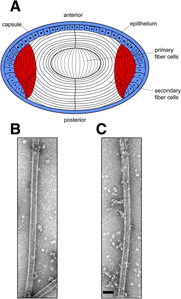
Figure 1. Preparation of microtubule associated proteins
A: Dissection of mouse lens epithelial and cortical fractions used for the isolation of microtubule associated proteins (MAPs). Wild type, αA-/--, αB-/--, and αA/B-/-- lenses were dissected into epithelial (blue) and cortical fiber cell (red) fractions. Microtubules were reconstituted from lens cortical fiber cell lysates and MAPs were extracted as described under Experimental Procedures. B and C: Electron micrographs of mouse lens cortical fiber microtubules that were used to dissociate the MAPs. Microtubules were negatively stained and examined. B shows wild type microtubules and C shows αB-/-- microtubules. The scale bar is equal to 50 nm.