![]() Table 1 of
Frost, Mol Vis 2007;
13:1580-1588.
Table 1 of
Frost, Mol Vis 2007;
13:1580-1588.
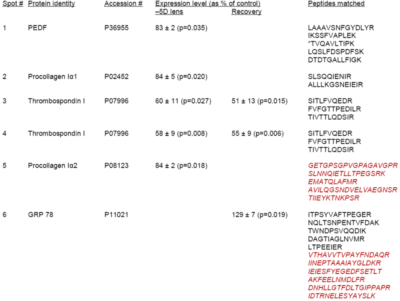
Table 1. Differentially expressed proteins identified from the scleral two-dimensional gels
Relative changes in expression as a percentage of the control (mean±SEM), and p-values, for proteins determined to be differentially expressed on the scleral 2D gels. Spots 1-5 were down-regulated in the treated eye during myopia development. During recovery from myopia spots 3 and 4 remained down-regulated while spots 1, 2, and 5 were no longer differentially expressed; spot 6 was up-regulated in the treated eye. Differentially expressed proteins were identified by mass spectrometry: peptides in black were determined by LC-MSMS, those in red italics by MALDI-TOF. All peptides matched human sequence entries in the Swiss-Prot database except for one (*) which matched the bovine sequence (GenBank Q95121).
