![]() Figure 4 of
Yzer, Mol Vis 2007;
13:1568-1572.
Figure 4 of
Yzer, Mol Vis 2007;
13:1568-1572.
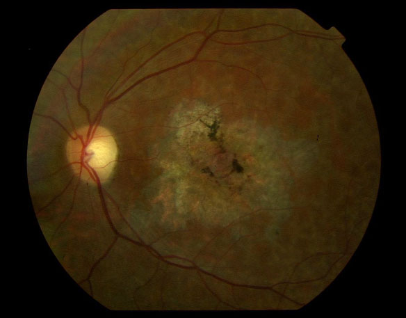
Figure 4. Fundus photograph of the left eye of patient VI:1, age 52
This photograph clearly shows the relatively normal optic disk, mild attenuation of the vessels, and large atrophic lesion with scattered pigmentations in the macula. The retinal pigment epithelium has a lobular atrophic appearance.