![]() Figure 3 of
Yzer, Mol Vis 2007;
13:1568-1572.
Figure 3 of
Yzer, Mol Vis 2007;
13:1568-1572.
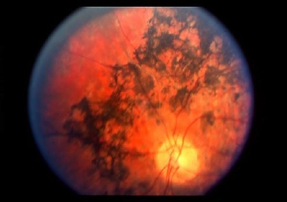
Figure 3. Fundus photograph of the right eye of patient V:5, age 43 years
Note the pale optic disk, moderate attenuation of the vessels, and heavy bone-spicule pigmentation in the midperiphery with a relatively spared periphery.