![]() Figure 3 of
Lambiase, Mol Vis 2007;
13:1562-1567.
Figure 3 of
Lambiase, Mol Vis 2007;
13:1562-1567.
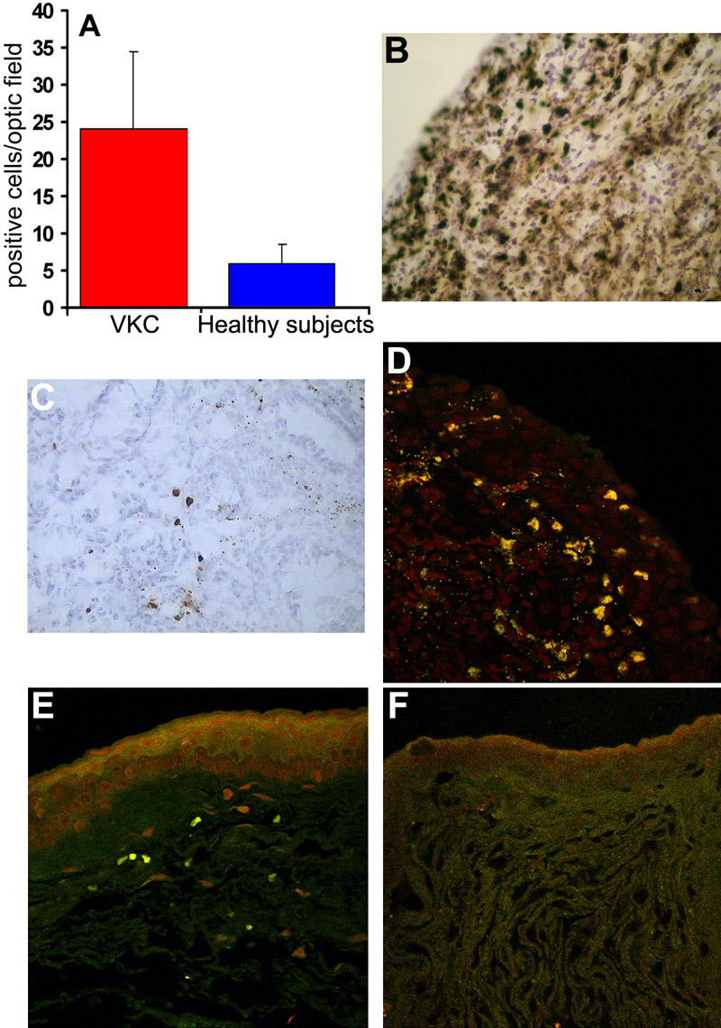
Figure 3. Immunohistochemical evaluation of natural killer cells in vernal keratoconjunctivitis conjunctiva
Graph A illustrates the significant increase of natural killer cells in the conjunctiva of patients with vernal keratoconjunctivitis (VKC) compared to healthy subjects. Natural killer cells were identified by CD56 expression quantified by immunohistochemistry (B=VKC and C=healthy subjects) and confocal analysis (D=VKC and E=healthy subjects). No specific immune staining was observed in the presence of isotype IgG (F=control staining; 40X magnification).