![]() Figure 2 of
Lambiase, Mol Vis 2007;
13:1562-1567.
Figure 2 of
Lambiase, Mol Vis 2007;
13:1562-1567.
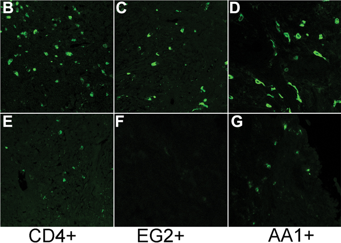
Figure 2. Evaluation of inflammatory cells in the conjunctiva of patients with vernal keratoconjunctivitis
The graph (A) illustrates a significant increase of T-helper lymphocytes (CD4+/green; B), activated eosinophils (EG2+/green; C), and mast cells (AA1+/green; D) in vernal keratoconjunctivitis (VKC) conjunctiva compared to healthy subjects (E, F, G, respectively). No specific binding was detected when sections were incubated with isotype IgG (data not shown; 40X magnification).