![]() Figure 4 of
Yuan, Mol Vis 2007;
13:1555-1561.
Figure 4 of
Yuan, Mol Vis 2007;
13:1555-1561.
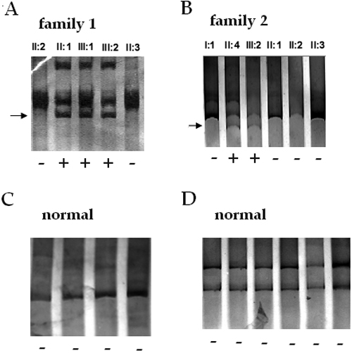
Figure 4. Detection of PAX6 mutations by single strand conformational polymorphism analysis
A: The exon 5 PCR product of PAX6 in Family 1 showed an extra band (arrow) on the second, third, and fourth lanes, which come from II:1, III:1, and III:2, respectively, on the 10% polyacrylamide gel, while the first and fifth lanes come from II:2 and II:3, which are unaffected families. B: The unusual banding pattern was observed in exon 9 of PAX6 in Family 2. The second and third lanes are II:4 and III:2; the other lanes are I:1, II:1, II:2, and II:3, which are the normals in Family 2. C and D: There is no unusual for the exon 5 and the exon 9 of control samples. A plus sign under a lane indicates a sample with a mutation and a minus sign indicates a normal.