![]() Figure 5 of
Ebermann, Mol Vis 2007;
13:1539-1547.
Figure 5 of
Ebermann, Mol Vis 2007;
13:1539-1547.
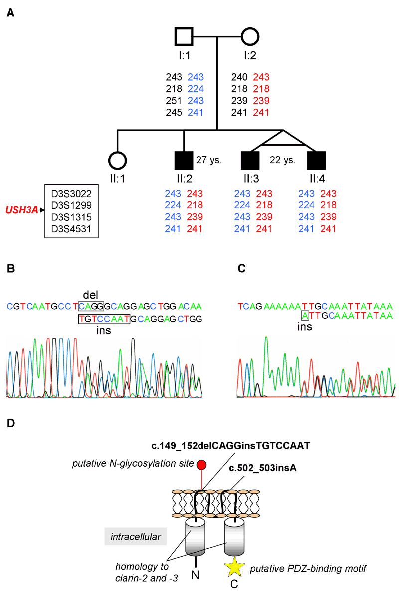
Figure 5. Genetic data from the study family
A: Pedigree of the German USH3 family investigated in this study. DNA was available from all persons displayed except II:1. Alleles for five microsatellite markers specific for the USH3A locus are given. The position of the USH3A gene relative to the markers is indicated. The three patients share the same haplotypes. B and C: Electropherograms for the heterozygous USH3A mutations c.149_152delCAGGinsTGTCCAAT and c.502_503insA, respectively. Deleted or inserted nucleotides are boxed. D: Drawing of the USH3A protein and position of the mutations identified in this study. In contrast to II.3, there is a significant affection of macular function in II:2, again demonstrating intrafamilial variability and showing that retinal degeneration can involve the center, and even at an early stage. The star designates the putative PDZ-binding motif.
