![]() Figure 2 of
Ebermann, Mol Vis 2007;
13:1539-1547.
Figure 2 of
Ebermann, Mol Vis 2007;
13:1539-1547.
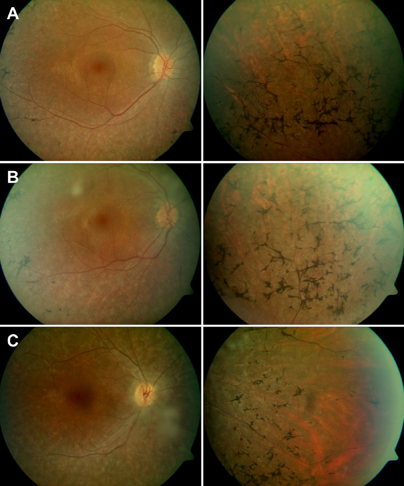
Figure 2. Fundus photographs from study patients
A: II:2 at 27 years of age, B: II:3 at 22 years of age, and C: II:4 at 22 years of age. Note slight waxy pallor of the optic disc (left column in A, B, and C), and thinning/loss of pigment epithelium with dense bone-spicule pigmentation in the middle periphery (right column in A, B, andC).