![]() Figure 7 of
Martins, Mol Vis 2007;
13:142-150.
Figure 7 of
Martins, Mol Vis 2007;
13:142-150.
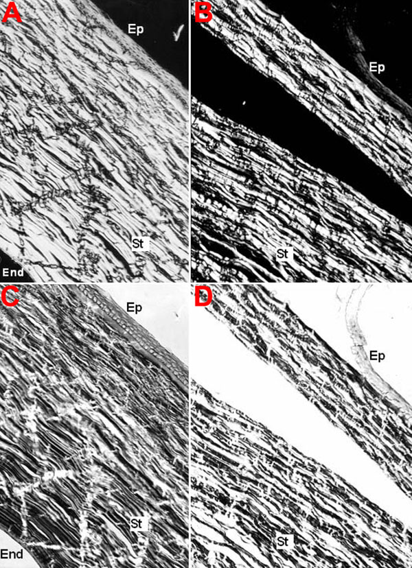
Figure 7. Control and LASIK human cornea birefringence images
The experiment was performed as describe in Methods, with (C,D) and without (A,B) compensation, for control (A,C) and LASIK-submitted (B,D) corneal slices. The samples are positioned 45° from the polarizer azimuth. Ep: epithelium; St: stroma; End: endothelium. The scale bar is equal to 20 μm.