![]() Figure 6 of
Martins, Mol Vis 2007;
13:142-150.
Figure 6 of
Martins, Mol Vis 2007;
13:142-150.
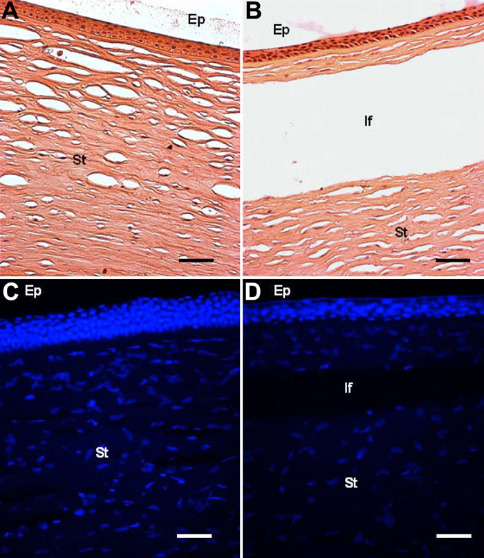
Figure 6. Hematoxilin and eosin staining and fluorescence microscopy for nuclei (DAPI) of normal and LASIK-treated corneal explants
The experiment was performed as described in Methods. Ep: epithelium; If: interface after LASIK; St: stroma. The scale bar equals 50 μm.