![]() Figure 5 of
Martins, Mol Vis 2007;
13:142-150.
Figure 5 of
Martins, Mol Vis 2007;
13:142-150.
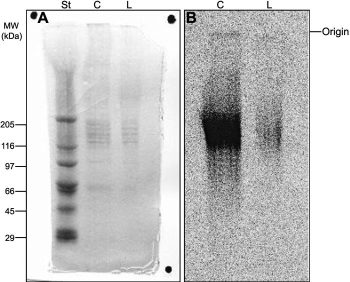
Figure 5. Polyacrylamide gel electrophoresis of proteoglycans extracted from human corneal explants
Aliquots (5 μl) of proteoglycans extracted from pair number 1 (see Table 1) of human corneal explants were submitted to polyacrylamide gel electrophoresis, as described in Methods. The gel was stained with coomassie blue (A), and the 35S-labeled compounds were localized by radioautography (B). St: molecular weight standard proteins; C: control cornea; L: LASIK submitted cornea.