![]() Figure 1 of
Martins, Mol Vis 2007;
13:142-150.
Figure 1 of
Martins, Mol Vis 2007;
13:142-150.
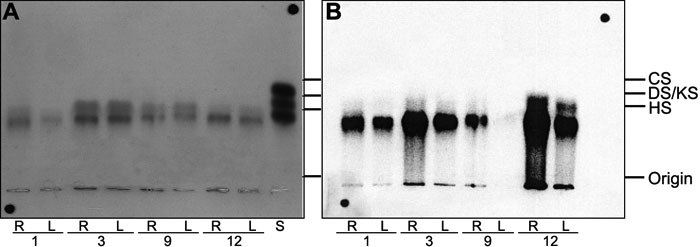
Figure 1. Agarose gel electrophoresis of proteoglycans extracted from human corneal explants
The left (L) cornea of each pair was submitted to LASIK, and the right (R) cornea was its matched control. The corneal explants were maintained under tissue culture conditions for 24 h in the presence of 35S-sulfate for the metabolic labeling of proteoglycans. The proteoglycans were extracted as described in Methods, and aliquots (5 μl) were submitted to agarose gel electrophoresis, as also described in Methods. The proteoglycans were fixed in the gel and stained by toluidine blue (A), and the 35S-labeled proteoglycans were localized by radioautography (B). In the images, S indicates a mixture of standard glycosaminoglycans containing chondroitin sulfate (CS), dermatan sulfate (DS), and heparan sulfate (HS), 5 μg each. KS indicates keratan sulfate. The numbers 1, 3, 9, and 12 refer to the corneal pairs described in Table 1.