![]() Figure 2 of
Bosley, Mol Vis 2007;
13:1516-1528.
Figure 2 of
Bosley, Mol Vis 2007;
13:1516-1528.
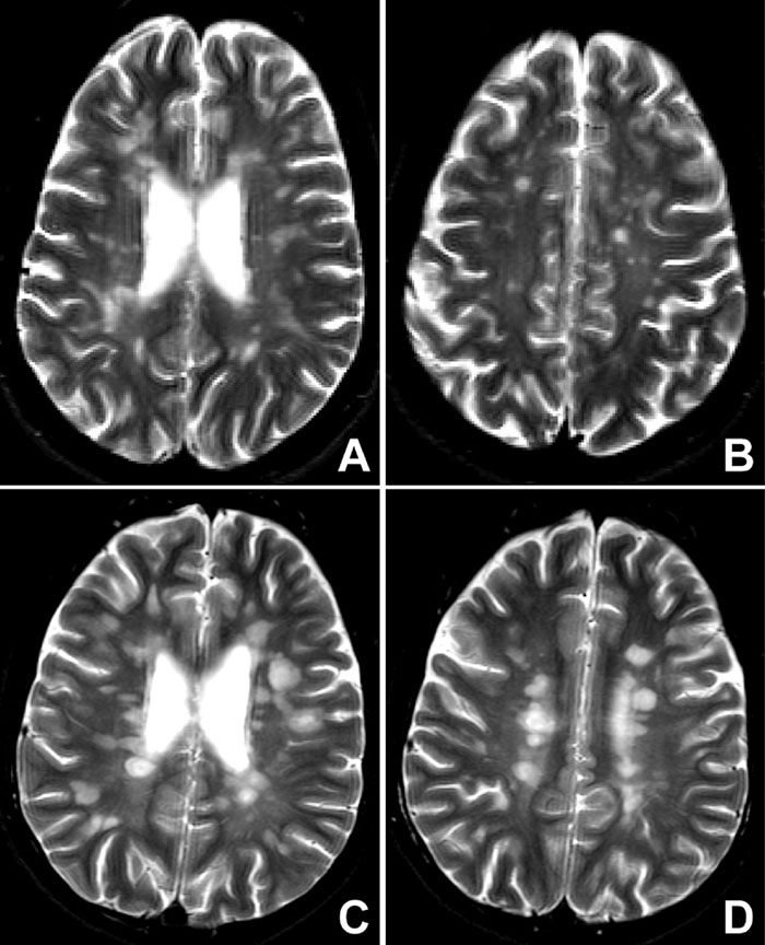
Figure 2. Disseminated demyelination in patients with optic neuritis
Montage of representative T2W MRI images of patients without (A and B; Patient 6) and with (C and D; Patient 16) potentially pathologic mitochondrial DNA changes showing a similar neuroimaging appearance typical of disseminated demyelinating disease that fulfills Fazekas criteria [14] despite the absence of clinically definite multiple sclerosis. MRI studies of optic neuritis patients showed a spectrum of demyelinating changes (see Table 1) that did not differ significantly according to mitochondrial parameters.