![]() Figure 1 of
Bosley, Mol Vis 2007;
13:1516-1528.
Figure 1 of
Bosley, Mol Vis 2007;
13:1516-1528.
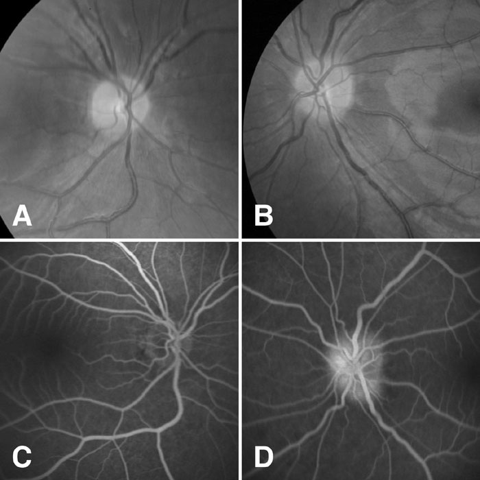
Figure 1. Mild papillitis in a patient with optic neuritis
(A) right eye (OD) and (B) left eye (OS) fundus photos of Patient 4, who had optic neuritis OS with mild swelling of the optic disc. Fluorescein angiogram OD (C; at 5:56 min) and OS (D; at 5:31 min) revealed staining of the left optic disc typical of papillitis but not compatible with the pseudoedema seen with Leber hereditary optic neuropathy [54].