![]() Figure 1 of
van Wijngaarden, Mol Vis 2007;
13:1508-1515.
Figure 1 of
van Wijngaarden, Mol Vis 2007;
13:1508-1515.
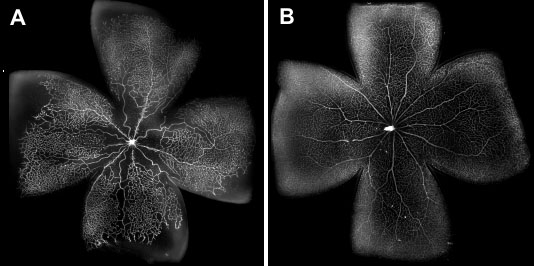
Figure 1. Montages of neonatal inbred Sprague Dawley rat retinal flat-mounts, labeled with fluorophore-conjugated isolectin GS-B4 to mark the retinal microvasculature
A: Shown are oxygen-induced retinopathy-affected retinal vasculature following 14 postnatal days of cyclic hyperoxic exposure. Note large avascular areas in the central and peripheral retina, and marked tortuosity of the major retinal vessels. B: Normal control retinal vasculature following 14 days exposure to the normoxia of room air. The retina is almost completely vascularized and shows no morphological abnormalities.