![]() Figure 8 of
Cristofanilli, Mol Vis 2007;
13:1496-1507.
Figure 8 of
Cristofanilli, Mol Vis 2007;
13:1496-1507.
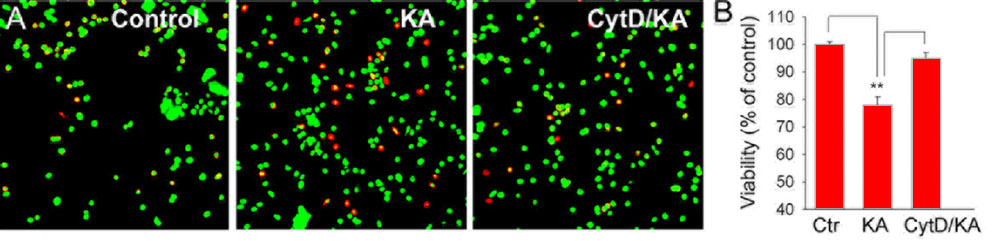
Figure 8. The effect of F-actin disruption on survival of retinal neurons in glutamate-induced excitotoxicity
Dissociated cell were incubated for 30 min in either normal Ringer solution A, B or Ringer solution containing 10 μM cytD (C) prior to exposure for 30 min to 100 μM kainate (B, C). Cells were then washed for 2 h and stained for live/dead assay. Calcein (green) stained healthy cells, whereas ethidium homodimer (red) stained dead cells. D: Cell viability was measured as the ratio of (live cells/total cells) x100, and normalized to control values (3 independent experiments).