![]() Figure 4 of
Cristofanilli, Mol Vis 2007;
13:1496-1507.
Figure 4 of
Cristofanilli, Mol Vis 2007;
13:1496-1507.
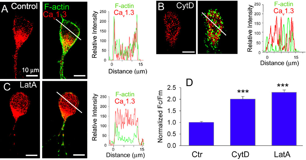
Figure 4. Depolymerization of F-actin causes Cav1.3 channel internalization in third-order retinal neurons
A: Subcellular localization of Cav1.3 channels (red) in third-order retinal neurons double stained with Alexa-Fluor488-phalloidin (green, middle panel). Intensity profiles show localization of Cav1.3 primarily on the surface membrane (right panel). B, C: Depolymerization of F-actin by either 10 μM cytD, or 5 μM latA reduced Cav1.3 labeling on the surface and increased it in the cytoplasm, as was evident from intensity profiles (right panels) obtained along the white lines. D: The bar graph summarizes effects of F-actin disrupters on internalization of Cav1.3 channel. Fc and Fm represent the cytoplasmic and the membrane immunofluorescence, respectively. Each column represents the mean±SE (n=30-50, 3-5 independent experiments, triple asterisks p<0.0001).