![]() Figure 1 of
Cristofanilli, Mol Vis 2007;
13:1496-1507.
Figure 1 of
Cristofanilli, Mol Vis 2007;
13:1496-1507.
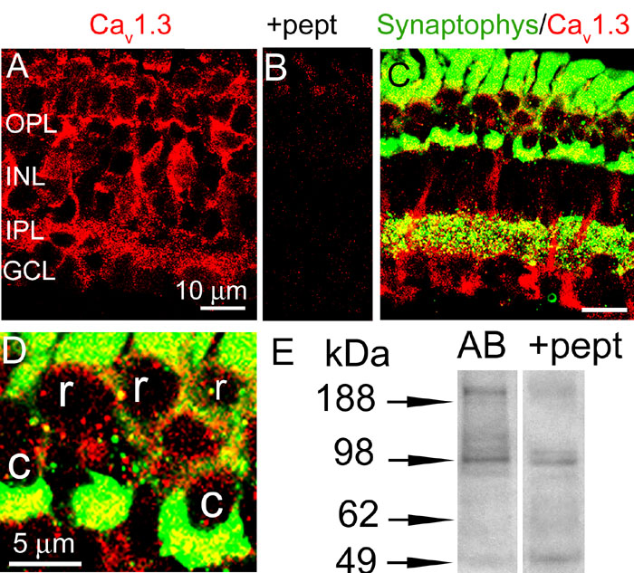
Figure 1. Localization of Cav1.3 L-type Ca channels in salamander retina
A: In salamander retinal slices Cav1.3 labeling was present in the outer plaxiform layer (OPL), inner plexiform layer (IPL) and on somas of cone photoreceptors and cells in the inner nuclear layer (INL), and ganglion cell layer (GCL). B: Preabsorbtion of antibody with respective peptide revealed only feint staining in photoreceptor inner segments. C: Double labeling with anti-synaptophysin (green) and anti-Cav1.3 (red) antibodies revealed aggregates of Cav1.3 puncta colocalized with synaptophysin in the OPL. Numerous puncta in the IPL also showed colocalization. D: Magnified image of OPL area shown in C, indicates localization of Cav1.3 channels in cone (c) photoreceptor terminals. E: In western blot analysis Cav1.3 antibody provided major bond around 200 kDa and a minor one around 140 kDa (n=4, 3 different retinal preparations). Preabsorption of anti- Cav1.3 with control peptide (+ pept) abolished the 200 kDa band.