![]() Figure 3 of
Scheef, Mol Vis 2007;
13:1483-1495.
Figure 3 of
Scheef, Mol Vis 2007;
13:1483-1495.
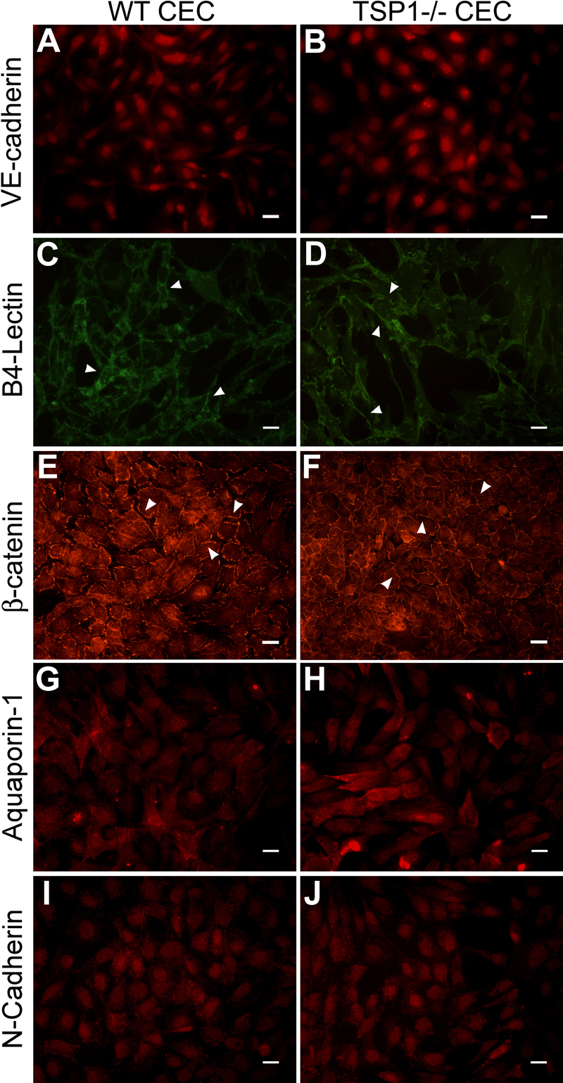
Figure 3. Localization of VE-cadherin, B4-lectin, β-catenin, aquaporin-1, and N-cadherin in CEC
Confluent monolayer of wild type (A, C, E, G, I) or TSP1-/- (B, D, F, H, J) CEC on glass coverslips were stained with anti-VE-cadherin (A, B), FITC-B4-lectin (C, D), anti-β-catenin (E, F), anti-aquaporin-1 (G, H), and anti-N-cadherin (I, J). Please note the junctional localization of B4-lectin and β-catenin (arrow heads), while other proteins showed defuse staining throughout the cells. These experiments were repeated at least twice with two different isolations of CEC. The scale baris equal to 20 μm.