![]() Figure 2 of
Scheef, Mol Vis 2007;
13:1483-1495.
Figure 2 of
Scheef, Mol Vis 2007;
13:1483-1495.
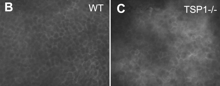
Figure 2. Expression of vascular endothelial cell markers in CEC
Mouse CEC were examined for expression of PECAM-1, VE-cadherin, and B4-lectin by FACS analysis (A). Please note that CEC express significant amounts of VE-cadherin and B4-lectin, but lack PECAM-1 expression. The shaded graphs show staining in the absence of primary antibody. B and C show corneal whole mounts prepared from wild type (B) and TSP1-/- (C) and stained with B4-lectin. Please note junctional localization of B4-lectin at sites of cell-cell contact.