![]() Figure 1 of
Scheef, Mol Vis 2007;
13:1483-1495.
Figure 1 of
Scheef, Mol Vis 2007;
13:1483-1495.
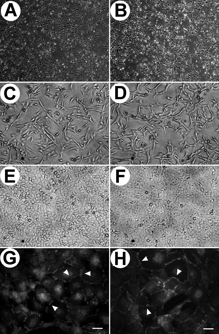
Figure 1. Morphology of mouse corneal endothelial cells (CEC) cultured on gelatin
Wild type CEC (A) and TSP1-/- CEC (B) were culture on gelatin-coated plates (x40). C and D are higher magnifications (x100) of A and B, respectively. E and F show morphology of cells at confluence (x100). G and H show ZO-1 staining. Please note the localization of ZO-1 to sites of cell-cell contact (arrow heads). The scale bar is equal to 20 μm.