![]() Figure 2 of
Lee, Mol Vis 2007;
13:1469-1474.
Figure 2 of
Lee, Mol Vis 2007;
13:1469-1474.
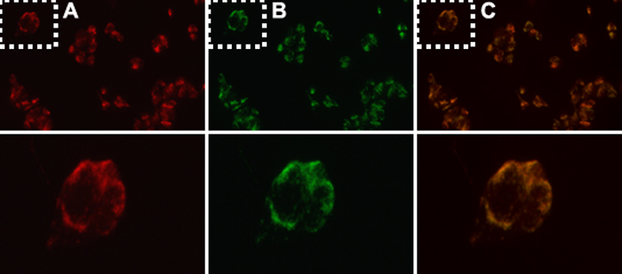
Figure 2. Mitochondrial colocalization of PRDX3 in human lens epithelial cells detected by immunofluoresence microscopy
Top panel, from left to right, represents: (A) Mitotracker alone (red), (B) PRDX3 (green), and (C) merging of the two images (orange). Bottom panel is a 60x magnification of the indicated regions from the top panel.