![]() Figure 4 of
Bagiyeva, Mol Vis 2007;
13:1458-1468.
Figure 4 of
Bagiyeva, Mol Vis 2007;
13:1458-1468.
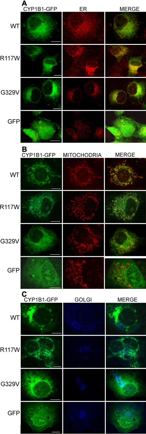
Figure 4. Subcellular localization of wild-type and R117W and G329V mutant CYP1B1 proteins by immunofluorescent confocal microscopy
The pEGFP-N2 empty vector was used as control. Note the non-specific localization of the GFP protein on its own, uniformly distributed within the cytosol and nucleoplasm. A: ER localization of CYP1B1 wild-type and mutants. In green, the CYP1B1-EGFP fusion protein; in red, anti-calnexinV. The merged images show colocalization in yellow. B: Mitochondrial localization of CYP1B1, particularly evident for the wild-type CYP1B1, but with less amounts of protein for the two, R117W and G329V, CYP1B1 mutants. In green, the CYP1B1-EGFP fusion protein; in red, Mitotracker. The merged images show colocalization in yellow. C: CYP1B1 is not localized in the Golgi in the wild-type and R117W CYP1B1 proteins, but there is some colocalization with the G329V mutant. In green, the CYP1B1-EGFP fusion protein; in bright blue, anti-GM130 (localized in the Golgi). The merged images show colocalization in light blue. The white bar indicates 10 μm size.