![]() Figure 3 of
Nakamura, Mol Vis 2007;
13:1451-1457.
Figure 3 of
Nakamura, Mol Vis 2007;
13:1451-1457.
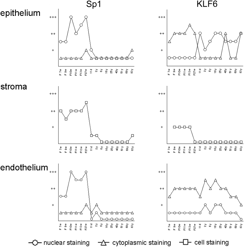
Figure 3. Line graphs summarizing immunostaining results for Sp1 and KLF6 in the developing cornea
The intensity of the staining in the nuclei (circles) and cytoplasm (triangles) of corneal cells was graded as undetectable (-), very weak (±), weak (+), moderate (++) or strong (+++). The Sp1 staining was mostly nuclear. The transition of cytoplasmic to nuclear staining for KLF6 was observed in the corneal epithelium and endothelium. In the corneal stroma, the intensity of total staining (squares) is shown, as it was difficult to discern whether the staining was nuclear or cytoplasmic.